What a fantastic exhibit of drawings at NYU’s Grey Gallery! Really astounding. Santiago Ramon y Cajal (1852-1934) studied art at an art academy as a teenager before going to medical school–his father, also in medicine, insisted on the change. Cajal’s drawing abilities–and more than that–his gorgeous linework, sometimes delicate and sometimes bold, tell a compelling story about how the brain works. He looked at the brain through a microscope, and then drew what he saw later, sometimes adding to drawings as he figured out more things about how the brain functioned. He was awarded the Nobel Prize in Physiology for asserting that “the brain is composed of individual neuron cells rather than a single continuous network.” (from the Grey Gallery exhibition text) A neurologist that was attending the exhibit today with a student entourage, told his group (I was eavesdropping) that the drawings Cajal made in the early 1900s are still used today by students of the brain.
The exhibit is worth seeing not only because of the many Cajal drawings, but also because there is artwork from previous eras, with alternate ideas of how the brain worked, and then modern, lovely MRI and photographic images of the brain that confirm the drawings Cajal made. What a pleasure to see it all together–and you can still get there! It closes March 31st.
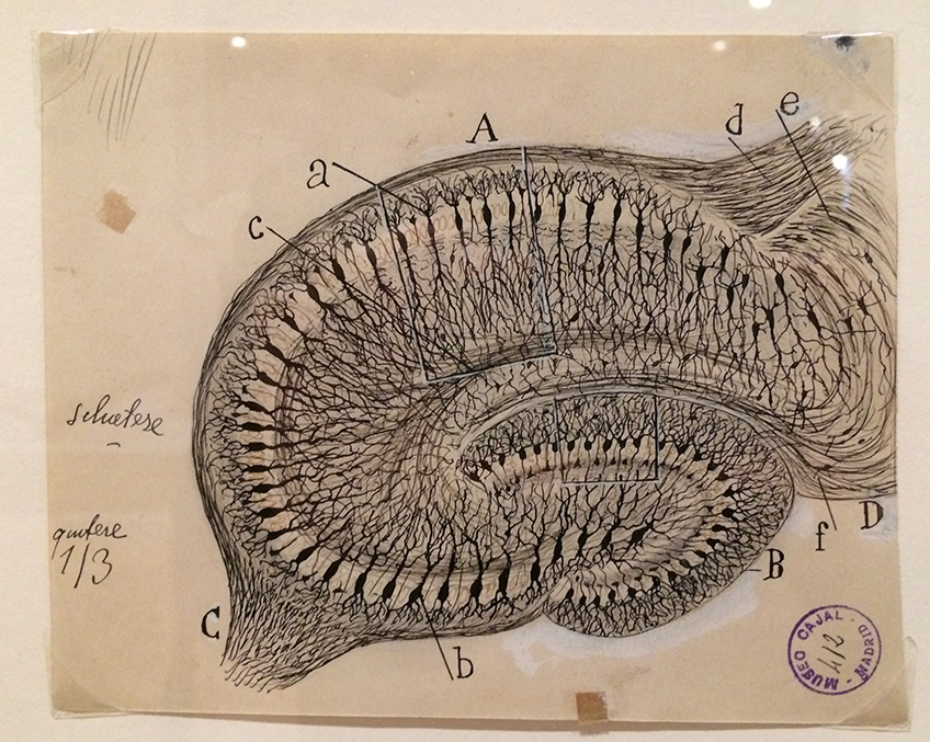
The Hippocampus, ink on paper
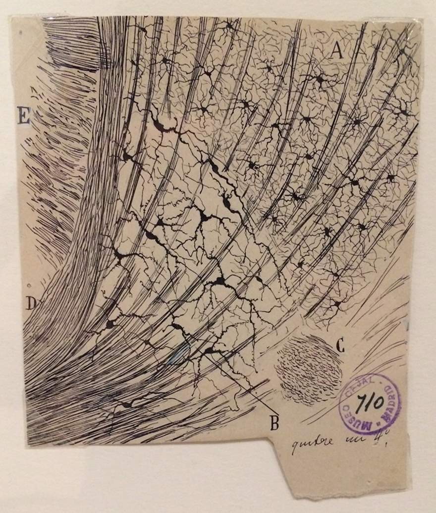
Caudate Nucleus of a twenty-day-old mouse (perhaps my favorite image, not sure why)
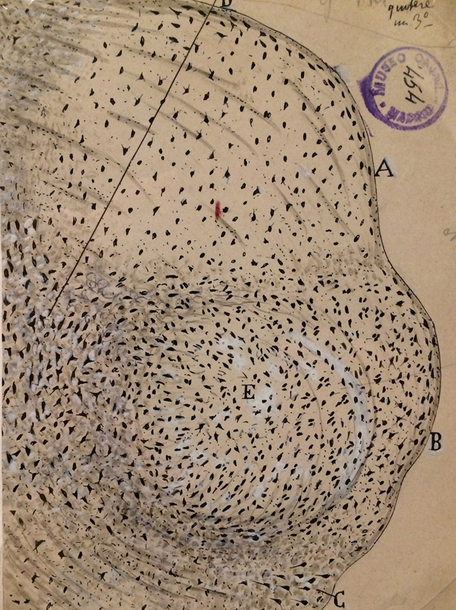
The medial geniculate nucleus in the thalamus of the cat, ink on paper
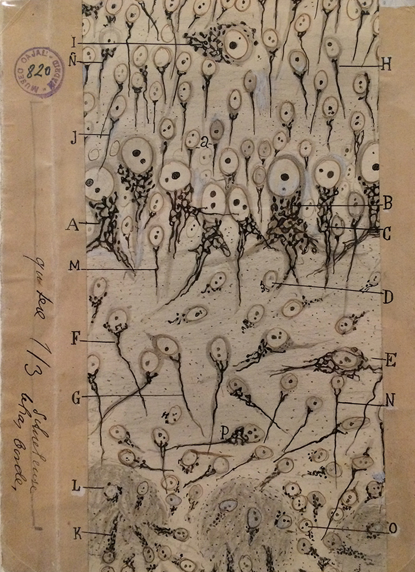
Hmm–I forgot to write down what this is. But–ink and pencil on paper certainly.
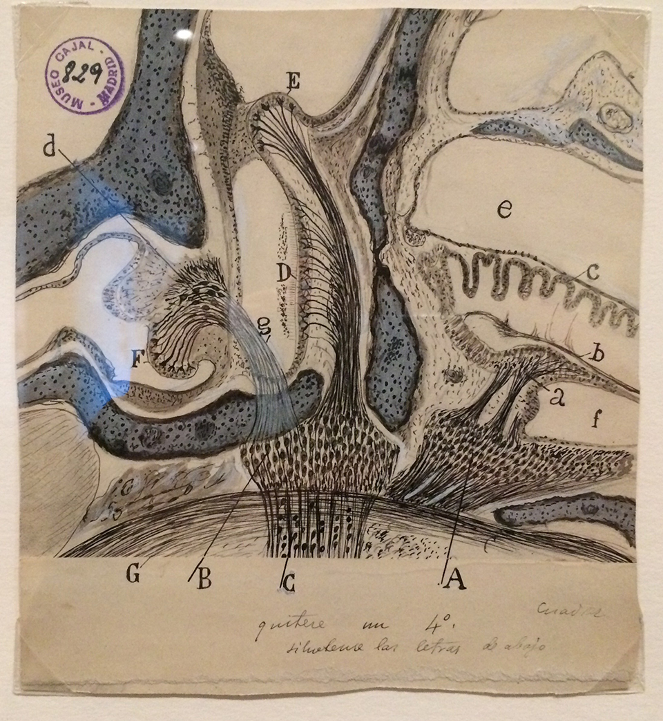
The labyrinth of the inner ear, ink and pencil on paper
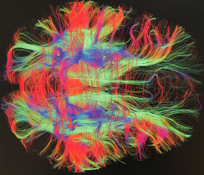
Zeynep M. Saygin: White matter fiber tracts of the brain in a healthy adult study participant, 2013, Diffusion tensor imaging MRI acquisition
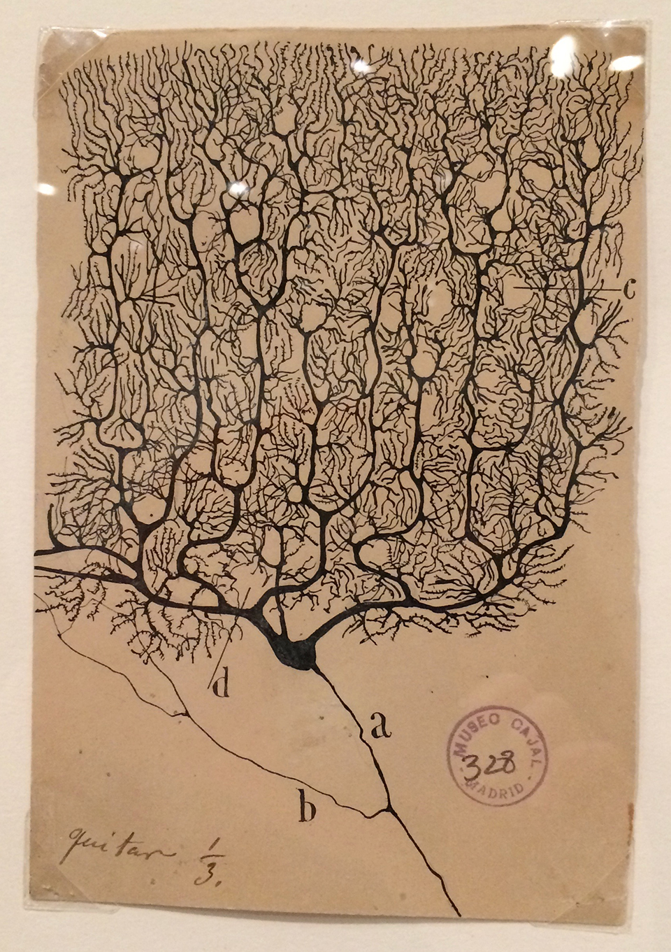
A Purkinje neuron from the human cerebellum, ink on paper
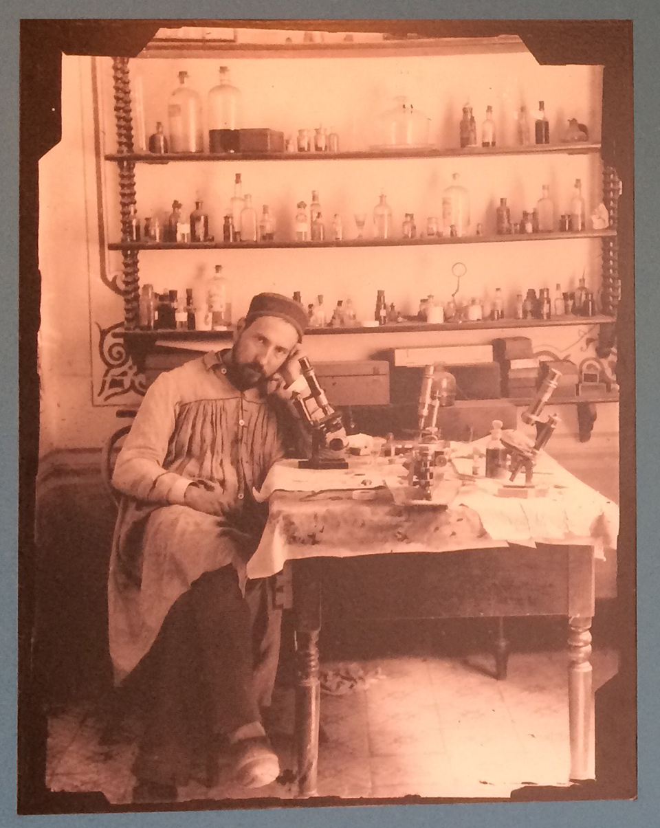
Santiago Ramon y Cajal in his laboratory/studio
